Why ser and not thr brokers catalysis in the trypsin fold

Pelc LA, Chen Z, Gohara DW, Vogt AD, Pozzi N, Di Cera E

Biochemistry 2015 Feb;54(7):1457-64

PMID: 25664608

Abstract

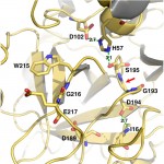
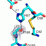
Although Thr is equally represented as Ser in the human genome and as a nucleophile is as good as Ser, it is never found in the active site of the large family of trypsin-like proteases that utilize the Asp/His/Ser triad. The molecular basis of the preference of Ser over Thr in the trypsin fold was investigated with X-ray structures of the thrombin mutant S195T free and bound to an irreversible active site inhibitor. In the free form, the methyl group of T195 is oriented toward the incoming substrate in a conformation seemingly incompatible with productive binding. In the bound form, the side chain of T195 is reoriented for efficient substrate acylation without causing steric clash within the active site. Rapid kinetics prove that this change is due to selection of an active conformation from a preexisting ensemble of reactive and unreactive rotamers whose relative distribution determines the level of activity of the protease. Consistent with these observations, the S195T substitution is associated with a weak yet finite activity that allows identification of an unanticipated important role for S195 as the end point of allosteric transduction in the trypsin fold. The S195T mutation abrogates the Na(+)-dependent enhancement of catalytic activity in thrombin, activated protein C, and factor Xa and significantly weakens the physiologically important allosteric effects of thrombomodulin on thrombin and of cofactor Va on factor Xa. The evolutionary selection of Ser over Thr in trypsin-like proteases was therefore driven by the need for high catalytic activity and efficient allosteric regulation.

Structural Dynamics as a Contributor to Error-prone Replication by an RNA-dependent RNA Polymerase

Moustafa IM, Korboukh VK, Arnold JJ, Smidansky ED, Marcotte LL, Gohara DW, Yang X, Sánchez-Farrán MA, Filman D, Maranas JK, Boehr DD, Hogle JM, Colina CM, Cameron CE

J. Biol. Chem. 2014 Dec;289(52):36229-48

PMID: 25378410

Abstract

RNA viruses encoding high- or low-fidelity RNA-dependent RNA polymerases (RdRp) are attenuated. The ability to predict residues of the RdRp required for faithful incorporation of nucleotides represents an essential step in any pipeline intended to exploit perturbed fidelity as the basis for rational design of vaccine candidates. We used x-ray crystallography, molecular dynamics simulations, NMR spectroscopy, and pre-steady-state kinetics to compare a mutator (H273R) RdRp from poliovirus to the wild-type (WT) enzyme. We show that the nucleotide-binding site toggles between the nucleotide binding-occluded and nucleotide binding-competent states. The conformational dynamics between these states were enhanced by binding to primed template RNA. For the WT, the occluded conformation was favored; for H273R, the competent conformation was favored. The resonance for Met-187 in our NMR spectra reported on the ability of the enzyme to check the correctness of the bound nucleotide. Kinetic experiments were consistent with the conformational dynamics contributing to the established pre-incorporation conformational change and fidelity checkpoint. For H273R, residues comprising the active site spent more time in the catalytically competent conformation and were more positively correlated than the WT. We propose that by linking the equilibrium between the binding-occluded and binding-competent conformations of the nucleotide-binding pocket and other active-site dynamics to the correctness of the bound nucleotide, faithful nucleotide incorporation is achieved. These studies underscore the need to apply multiple biophysical and biochemical approaches to the elucidation of the physical basis for polymerase fidelity.

Sequence selectivity of macrolide-induced translational attenuation

Davis AR, Gohara DW, Yap MN

Proc. Natl. Acad. Sci. U.S.A. 2014 Oct;111(43):15379-84

PMID: 25313041

Abstract

The prevailing “plug-in-the-bottle” model suggests that macrolide antibiotics inhibit translation by binding inside the ribosome tunnel and indiscriminately arresting the elongation of every nascent polypeptide after the synthesis of six to eight amino acids. To test this model, we performed a genome-wide analysis of translation in azithromycin-treated Staphylococcus aureus. In contrast to earlier predictions, we found that the macrolide does not preferentially induce ribosome stalling near the 5′ end of mRNAs, but rather acts at specific stalling sites that are scattered throughout the entire coding region. These sites are highly enriched in prolines and charged residues and are strikingly similar to other ligand-independent ribosome stalling motifs. Interestingly, the addition of structurally related macrolides had dramatically different effects on stalling efficiency. Our data suggest that ribosome stalling can occur at a surprisingly large number of low-complexity motifs in a fashion that depends only on a few arrest-inducing residues and the presence of a small molecule inducer.

Crystal structure of prothrombin reveals conformational flexibility and mechanism of activation

Pozzi N, Chen Z, Gohara DW, Niu W, Heyduk T, Di Cera E

J. Biol. Chem. 2013 Aug;288(31):22734-44

PMID: 23775088

Abstract

The zymogen prothrombin is composed of fragment 1 containing a Gla domain and kringle-1, fragment 2 containing kringle-2, and a protease domain containing A and B chains. The prothrombinase complex assembled on the surface of platelets converts prothrombin to thrombin by cleaving at Arg-271 and Arg-320. The three-dimensional architecture of prothrombin and the molecular basis of its activation remain elusive. Here we report the first x-ray crystal structure of prothrombin as a Gla-domainless construct carrying an Ala replacement of the catalytic Ser-525. Prothrombin features a conformation 80 Å long, with fragment 1 positioned at a 36° angle relative to the main axis of fragment 2 coaxial to the protease domain. High flexibility of the linker connecting the two kringles suggests multiple arrangements for kringle-1 relative to the rest of the prothrombin molecule. Luminescence resonance energy transfer measurements detect two distinct conformations of prothrombin in solution, in a 3:2 ratio, with the distance between the two kringles either fully extended (54 ± 2 Å) or partially collapsed (≤34 Å) as seen in the crystal structure. A molecular mechanism of prothrombin activation emerges from the structure. Of the two sites of cleavage, Arg-271 is located in a disordered region connecting kringle-2 to the A chain, but Arg-320 is well defined within the activation domain and is not accessible to proteolysis in solution. Burial of Arg-320 prevents prothrombin autoactivation and directs prothrombinase to cleave at Arg-271 first. Reversal of the local electrostatic potential then redirects prothrombinase toward Arg-320, leading to thrombin generation via the prethrombin-2 intermediate.

Cohesin and polycomb proteins functionally interact to control transcription at silenced and active genes

Schaaf CA, Misulovin Z, Gause M, Koenig A, Gohara DW, Watson A, Dorsett D

PLoS Genet. 2013 Jun;9(6):e1003560

PMID: 23818863

Abstract

Cohesin is crucial for proper chromosome segregation but also regulates gene transcription and organism development by poorly understood mechanisms. Using genome-wide assays in Drosophila developing wings and cultured cells, we find that cohesin functionally interacts with Polycomb group (PcG) silencing proteins at both silenced and active genes. Cohesin unexpectedly facilitates binding of Polycomb Repressive Complex 1 (PRC1) to many active genes, but their binding is mutually antagonistic at silenced genes. PRC1 depletion decreases phosphorylated RNA polymerase II and mRNA at many active genes but increases them at silenced genes. Depletion of cohesin reduces long-range interactions between Polycomb Response Elements in the invected-engrailed gene complex where it represses transcription. These studies reveal a previously unrecognized role for PRC1 in facilitating productive gene transcription and provide new insights into how cohesin and PRC1 control development.

Genome-wide control of RNA polymerase II activity by cohesin

Schaaf CA, Kwak H, Koenig A, Misulovin Z, Gohara DW, Watson A, Zhou Y, Lis JT, Dorsett D

PLoS Genet. 2013 Mar;9(3):e1003382

PMID: 23555293

Abstract

Cohesin is a well-known mediator of sister chromatid cohesion, but it also influences gene expression and development. These non-canonical roles of cohesin are not well understood, but are vital: gene expression and development are altered by modest changes in cohesin function that do not disrupt chromatid cohesion. To clarify cohesin’s roles in transcription, we measured how cohesin controls RNA polymerase II (Pol II) activity by genome-wide chromatin immunoprecipitation and precision global run-on sequencing. On average, cohesin-binding genes have more transcriptionally active Pol II and promoter-proximal Pol II pausing than non-binding genes, and are more efficient, producing higher steady state levels of mRNA per transcribing Pol II complex. Cohesin depletion frequently decreases gene body transcription but increases pausing at cohesin-binding genes, indicating that cohesin often facilitates transition of paused Pol II to elongation. In many cases, this likely reflects a role for cohesin in transcriptional enhancer function. Strikingly, more than 95% of predicted extragenic enhancers bind cohesin, and cohesin depletion can reduce their association with Pol II, indicating that cohesin facilitates enhancer-promoter contact. Cohesin depletion decreases the levels of transcriptionally engaged Pol II at the promoters of most genes that don’t bind cohesin, suggesting that cohesin controls expression of one or more broadly acting general transcription factors. The multiple transcriptional roles of cohesin revealed by these studies likely underlie the growth and developmental deficits caused by minor changes in cohesin activity.

Conformational selection in trypsin-like proteases

Pozzi N, Vogt AD, Gohara DW, Di Cera E

Curr. Opin. Struct. Biol. 2012 Aug;22(4):421-31

PMID: 22664096

Abstract

For over four decades, two competing mechanisms of ligand recognition–conformational selection and induced-fit–have dominated our interpretation of protein allostery. Defining the mechanism broadens our understanding of the system and impacts our ability to design effective drugs and new therapeutics. Recent kinetics studies demonstrate that trypsin-like proteases exist in equilibrium between two forms: one fully accessible to substrate (E) and the other with the active site occluded (E*). Analysis of the structural database confirms existence of the E* and E forms and vouches for the allosteric nature of the trypsin fold. Allostery in terms of conformational selection establishes an important paradigm in the protease field and enables protein engineers to expand the repertoire of proteases as therapeutics.

Genome-wide networks of amino acid covariances are common among viruses

Donlin MJ, Szeto B, Gohara DW, Aurora R, Tavis JE

J. Virol. 2012 Mar;86(6):3050-63

PMID: 22238298

Abstract

Coordinated variation among positions in amino acid sequence alignments can reveal genetic dependencies at noncontiguous positions, but methods to assess these interactions are incompletely developed. Previously, we found genome-wide networks of covarying residue positions in the hepatitis C virus genome (R. Aurora, M. J. Donlin, N. A. Cannon, and J. E. Tavis, J. Clin. Invest. 119:225-236, 2009). Here, we asked whether such networks are present in a diverse set of viruses and, if so, what they may imply about viral biology. Viral sequences were obtained for 16 viruses in 13 species from 9 families. The entire viral coding potential for each virus was aligned, all possible amino acid covariances were identified using the observed-minus-expected-squared algorithm at a false-discovery rate of ≤1%, and networks of covariances were assessed using standard methods. Covariances that spanned the viral coding potential were common in all viruses. In all cases, the covariances formed a single network that contained essentially all of the covariances. The hepatitis C virus networks had hub-and-spoke topologies, but all other networks had random topologies with an unusually large number of highly connected nodes. These results indicate that genome-wide networks of genetic associations and the coordinated evolution they imply are very common in viral genomes, that the networks rarely have the hub-and-spoke topology that dominates other biological networks, and that network topologies can vary substantially even within a given viral group. Five examples with hepatitis B virus and poliovirus are presented to illustrate how covariance network analysis can lead to inferences about viral biology.

Allostery in trypsin-like proteases suggests new therapeutic strategies

Gohara DW, Di Cera E

Trends Biotechnol. 2011 Nov;29(11):577-85

PMID: 21726912

Abstract

Trypsin-like proteases (TLPs) are a large family of enzymes responsible for digestion, blood coagulation, fibrinolysis, development, fertilization, apoptosis and immunity. A current paradigm posits that the irreversible transition from an inactive zymogen to the active protease form enables productive interaction with substrate and catalysis. Analysis of the entire structural database reveals two distinct conformations of the active site: one fully accessible to substrate (E) and the other occluded by the collapse of a specific segment (E*). The allosteric E*-E equilibrium provides a reversible mechanism for activity and regulation in addition to the irreversible zymogen to protease conversion and points to new therapeutic strategies aimed at inhibiting or activating the enzyme. In this review, we discuss relevant examples, with emphasis on the rational engineering of anticoagulant thrombin mutants.